Introduction:
Allogeneic hematopoietic cell transplantation (HCT) is potentially curative for patients with sickle cell disease (SCD). It is often recommended for patients with severe SCD, including those at risk of, or with a history of stroke. There has been limited investigation into the direct impact of HCT on cerebral hemodynamics. Quantitative biomarkers of cerebral hemodynamics, including oxygen extraction fraction (OEF) and cerebral blood flow (CBF), can provide an objective assessment of therapeutic efficacy of HCT as well as other emerging therapies. We have previously shown that CBF improves after HCT in patients with SCD (Sharma A, et al. Suppl 1, Blood 2021). Here, we performed a prospective evaluation using magnetic resonance imaging (MRI) to measure the OEF of the whole brain and the superior sagittal sinus (SSS), to compare cerebral oxygen metabolism in SCD patients before and after HCT.
Methods:
We performed anatomical and hemodynamic MRI of the brain prior to, and at 6 months after HCT in children with SCD undergoing a reduced intensity conditioning based HCT on a clinical trial (NCT04362293). Patients were receiving either hydroxyurea (HU) or chronic transfusion therapy (CTT) before HCT. MRI brain scanning was performed for all patients in a 3 Tesla MRI imaging system (Siemens MAGNETOM Prisma, Erlangen, Germany) with a 16-channel phased-array coil. Quantitative susceptibility map (QSM) imaging was performed through a 3D multi-echo gradient echo sequence with flow compensation and reconstructed with the morphology-enabled dipole inversion (MEDI) method (Liu T, et al. MRM 2013). The oxygen extraction fraction was calculated utilizing the established formula incorporating clinically measured hematocrit, tissue susceptibility, and hemoglobin susceptibility values quantified through quantitative susceptibility mapping (QSM), as previously delineated (Fan A, et al. JCBFM 2020) for the segmented vessels from the whole brain and the SSS.
Results:
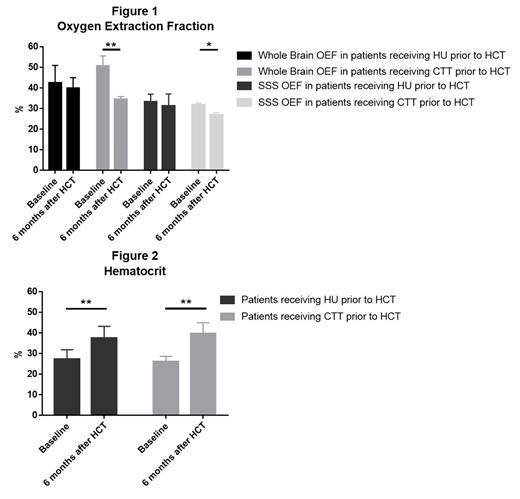
The median age at HCT was 15.6 years (N=11). Hematocrit as well as global and regional (SSS) OEF of the patients were quantified and compared between baseline and at 6 months after HCT (Fig 1). All patients experienced a decrease in OEF after HCT. We then stratified the patients into those who were receiving HU or CTT prior to HCT. In patients receiving HU prior to HCT (n=7), there was a small decrease in OEF for the whole brain (42.4% vs. 39.9%, P = 0.40) and SSS (33.1% vs. 31.3%, P =0.12) at 6 months after HCT compared to baseline. However, in patients receiving CTT prior to HCT (n=4) both the global OEF (50.0% vs. 34.4 %, P < 0.01) and regional OEF (31.8% vs. 26.7 %, P < 0.05) decreased significantly at 6 months after HCT. All patients exhibited an increased hematocrit following HCT (Fig 2). Specifically, the HU cohort improved from a mean of 27.4% to 37.5% ( P<0.01), while the CTT cohort increased from 26.2% to 39.6% ( P<0.01) after HCT.
Conclusions:
Our results demonstrate the feasibility of global and regional OEF quantification using MRI in patients undergoing HCT for SCD. We observed that the OEF of the whole brain and SSS exhibited a similar downward trend after HCT in patients receiving either HU or CTT. This indicates an overall improvement in oxygen delivery after HCT. OEF decrease was marked after HCT in the CTT group, suggesting that HCT provided better cerebral hemodynamic outcomes than at baseline for this group. In contrast, the cohort of patients receiving HU showed a subtle decrease in OEF after HCT, perhaps implying that these patients had milder cerebral hemodynamic compromise than the CTT group. Both pretreatment groups demonstrated significantly higher hemoglobin concentrations after HCT compared to before treatment which may have contributed to the improvement in OEF.
Our study presents objective physiologic comparisons of different clinical therapies on cerebral hemodynamics, providing early data on the effects of cerebral oxygen delivery in pediatric patients with SCD. Further studies with larger patient numbers are warranted to confirm the trends observed in oxygen metabolism and understand the mechanisms underlying differential responses to HCT and other disease-modifying therapies. These imaging biomarkers may also help assess different genetic therapies currently in development.
Disclosures
Sharma:Vertex Pharmaceuticals: Consultancy, Other: Clinical Trial Site PI; RCI BMT/NMDP: Honoraria, Other: Clinical Trial Medical Monitor; CRISPR Therapeutics: Other: Clinical Trial Site PI, Research Funding; Editas Medicine: Consultancy; Sangamo Therapeutics: Consultancy; Medexus Inc: Consultancy.


